
Retinal vasculitis and cystoid macular odema vitreous cells
740
| Wegeners Granulomatosis – Itis a granulomatous necrotizing vasculitic condition that primarily affects upper and lower respiratory tracts and kidneys. In addition to retinal vasculitis, other clinical findings | ||||
|---|---|---|---|---|
| include | necrotizing | scleritis, | ||
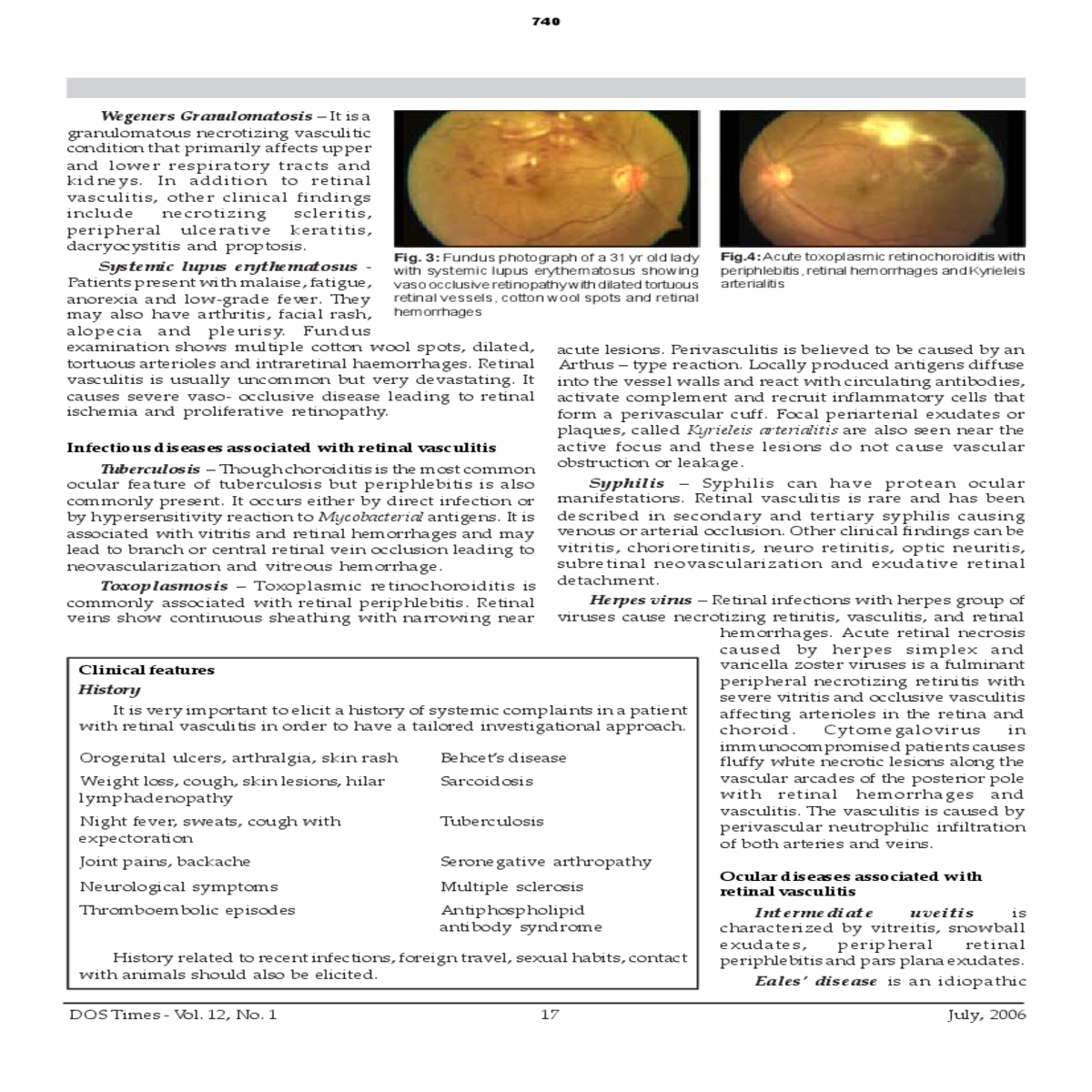
| Fig. 3: Fundus photograph of a 31 yr old lady | ||||
| with systemic lupus erythematosus showing | ||||
| vaso occlusive retinopathy with dilated tortuous |
|
|||
|
|---|
Tuberculosis – Though choroiditis is the most common ocular feature of tuberculosis but periphlebitis is also commonly present. It occurs either by direct infection or by hypersensitivity reaction to Mycobacterial antigens. It is associated with vitritis and retinal hemorrhages and may lead to branch or central retinal vein occlusion leading to neovascularization and vitreous hemorrhage.
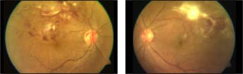
acute lesions. Perivasculitis is believed to be caused by an Arthus – type reaction. Locally produced antigens diffuse into the vessel walls and react with circulating antibodies, activate complement and recruit inflammatory cells that form a perivascular cuff. Focal periarterial exudates or plaques, called Kyrieleis arterialitis are also seen near the active focus and these lesions do not cause vascular obstruction or leakage.
caused by herpes simplex and
|
|||||||
|---|---|---|---|---|---|---|---|
| choroid. | Cytomegalovirus | in | |||||
|
|||||||
| Intermediate | uveitis | is | |||||
|
|||||||
|
17 | July, 2006 | |||||
741
 |
||||
|---|---|---|---|---|
| alterations | and | |||
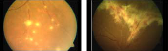
| Fig.5: Fundus photograph showing multiple skip | Fig.6: Fundus photograph of a patient with | |||
| lesions of vascular cuffing (taches de bougie) | cytomegalovirus retinitis showing fluffy white | |||
|
|
|
|---|---|---|
Birdshot retinochoroidopathy is a bilateral panuveitis where fundus examination shows cream colored, deep, round lesions, retinal vasculitis and cystoid macular odema.
aqueous or vitreous cells, aqueous flare, keratic precipitates, posterior synechiae and posterior subcapsular cataract.
| reported | in | herpes, | rubella, | ||
|---|---|---|---|---|---|
|
|||||
| 18 | DOS Times - Vol. 12, No. 1 | ||||


