

Pelvic girdle and the lower limbs
Special Needs of A Mobile and Theatre Modality in
Assessing and Diagnosis
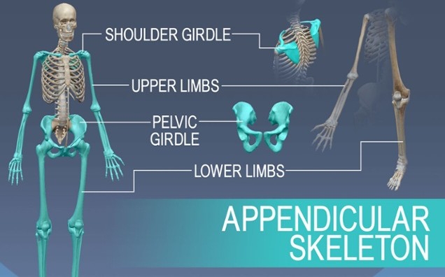
Bones of the human skeleton are divided into two main groups including axial skeleton and perpendicular skeleton. The bones of the appendicular skeleton creates the remaining skeleton. These are called appendicular because they are attachments of the axial skeleton. The appendicular skeleton comprises of the bones of the shoulder girdle, upper limbs, pelvic girdle and the lower limbs. Old-styled appendicular skeleton X-rays can help your doctor classify any damage from a shocking fall or mishap observe the development of a disease or trail the effects of assured treatment methods. Skeletal mobile X-rays are often done on an emergency basis after a suffering, such as a fall or misfortune. The physician will order an X-ray in any area that is causing immense pain in order to govern whether you have one or more broken bones. The physician may order a skeletal mobile X-ray if you display any marks or symptoms of circumstances that affect the bones, such as pain or swelling. These involve: arthritis, bone cancer, cancer that has spread to the bone, fractures, infections, osteoporosis and dental conditions. Contingent to the part under evaluation, the patient has to wear loose, contented clothing so that it is tranquil to move around. The patient is asked to take off any jewelry, spectacles, earsplitting or other metal items from your body before the mobile X-ray. The patient should inform the doctor if he/she have any metal grafts from prior surgeries. These may include a heart valve or pacemaker. In some occurrences the doctor may have selected to order a mobile X-ray because the patient have metal entrenched in the body. Different scans including MRI, can be dangerous for people with metal grafts.[Ske05]
4. Describe common types of surgical procedures that are performed with c-arm Mobile C-arm mobile X-ray systemsare used for different types of diagnostic imaging and nominally intrusive surgical procedures. In the operating room they aid in imagining kidney drainage, abdominal and thoracic aortic aneurysm reconstruction, percutaneous valve alternatives, cardiac surgery, vascular surgery, gastroenterology, NEUROspur, orthopedics, management of pain and neurology processes. Mobile C-arm includes a category of mini C-arms that are tiny systems used in clinics for sports remedy, bone operations and podiatric imaging. Latest specifications of modern mobile C-arm systems include 3-D navigation services and image processing software to enhance image eminence and system competences. The cardiac and vascular mockups of GE/OEC imaging structures use motion accepting subtraction progressive processing procedures to allow real-time subtraction without using a mask image. This enables the physician to perform a complete limit run-off with a solitary contrast media inoculation and a single imaging run. Many systems propose technologies to decrease the radiation dose supplied to patients. Other specifications involve a touch-screen interface, laser pointing guides, digital subtraction angiograph and process road mapping, permitting vascular processes to be premeditated with least amounts of distinction media and smaller fluoroscopy times.[AnI11]
Works Cited
An Introduction to Mobile C-Arm X-Ray Systems. (2011). Retrieved from itnonline: https://www.itnonline.com/article/introduction-mobile-c-arm-x-ray-systems
| Trauma | Imaging. | (2015). | Retrieved | from |
|---|
https://emedicine.medscape.com/article/357007-overview


