
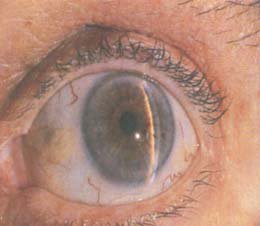
Ocular intermittent angle closure glaucoma
940
 |
 |
 |
|---|
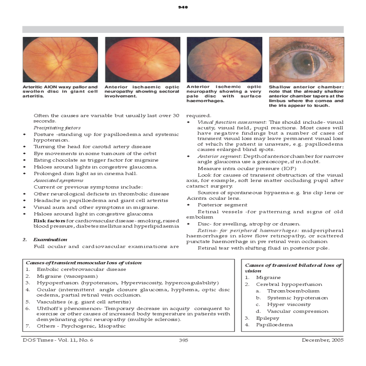
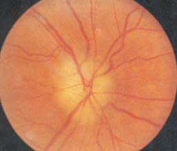
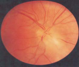
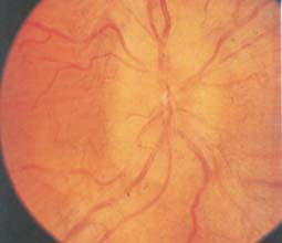
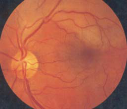
| Arteritic AION waxy pallor and swollen disc in giant cell | ischemic | optic | |||||||||||
|---|---|---|---|---|---|---|---|---|---|---|---|---|---|
|
involvement. |
|
surface | ||||||||||
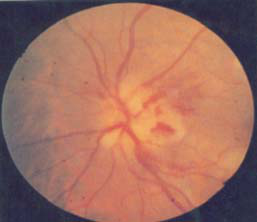
| the iris appear to touch. | |||||||||||||
|
� |
|
|||||||||||
| � | |||||||||||||
|
|
||||||||||||
| � | � | ||||||||||||
| � | |||||||||||||
|
|||||||||||||
| � | |||||||||||||
|
|||||||||||||
| � | |||||||||||||
| � |
|
||||||||||||
|
|||||||||||||
| � | Other neurological deficiets in thrombolic disease | ||||||||||||
| � |
|
|
|||||||||||
| � | � | ||||||||||||
| � | |||||||||||||
|
|||||||||||||
|
|||||||||||||
| � | |||||||||||||
| Retina- for peripheral haemorrhages: midperipheral | |||||||||||||
| 2. | |||||||||||||
|
|||||||||||||
|
|||||||||||||
|
|
||||||||||||
|
|
December, 2005 | |||||||||||
|
 |
||
|---|---|---|---|
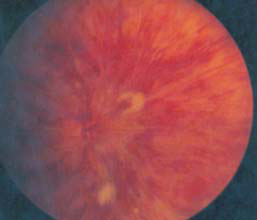
| Central retinal vein occlusion (ischaemic) showing multiple | |||
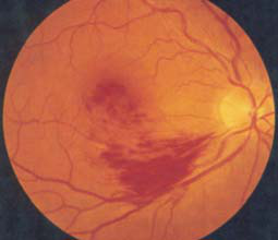
| showing cotton wool spot haemorrhage from disc vessel | cilioretinal sparing: emboli at the first bifurcation of the | ||
| margins are blurred, the | |||
| in a patient with almost complete obstruction of both | temporal branch of the central retinal artery and more | vessels emerging from the disc are partly buried, and the | |
| carotids. | capillaries on the nerve head | ||
|
 |
 |
 |
||||||||
|---|---|---|---|---|---|---|---|---|---|---|
| Branch retinal vein occlusion | retinal | artery | Branch | retinal | artery | |||||
|
|
|||||||||
| narrowed arterioles. | ||||||||||
|
Cardiovascular system: Pulse and rhythm, BP, carotid | |||||||||
|
||||||||||
| 3. | ||
|---|---|---|
| The ESR should be measured for all patients. A high ESR |
in patients over the age of 50 strongly suggests giant cell
|
B) | |
|---|---|---|
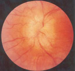
| � | Anterior ischemic neuropathy (AION) is caused by |
considered. Signs and symptoms- There is sudden loss of vision
and swelling of optic disc. Two forms of AION are
| A) | � |
|
|||
|
|||||
|
|
DOS Times - Vol. 11, No. 6 | |||


