Language:EN
Pages: 20
Words: 2911
Rating : ⭐⭐⭐⭐⭐
Price: $10.99
Page 1 Preview
incomplete merging the maxillary and mandibular pr

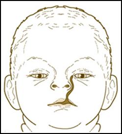
Incomplete merging the maxillary and mandibular processes

Craniofacial Development

I.  Pharyngeal apparatus

  • pharyngeal arches are paired structures associated with the pharynx that contribute greatly to the formation of the face, jaw, ear, and neck

      • neural crest mesenchyme develops into bonecartilage, and/or connective tissue in each arch.

    • each pharyngeal arch has a cranial nerve associated with it:

      • arch 6: CN X (recurrent laryngeal branch of the vagus)

    • the inside of the pharyngeal apparatus is lined by endoderm that forms infoldings or pouches between the arches; since there are 5 pharyngeal arches, there are 4 pharyngeal pouches, the fates of which are discussed below.

A. Fates of pharyngeal clefts

The pharyngeal clefts are ectodermal-lined recesses that appear on the OUTSIDE of the pharnyx between the arches; cleft 1 is between arch 1 and 2, cleft 2 is between arches 2 and 3, etc.


  • splits into an upper maxillary prominence and a lower mandibular prominence

  • derivatives of the 1st arch are:

  • innervated by CN VII (facial nerve)

  • derivatives of the 2nd arch are:

  • innervated by CN IX (glossopharyngeal nerve)

  • derivatives of the 3rd arch are:

  • innervated by CN X (superior laryngeal branch of the vagus nerve)

  • derivatives of the 4th arch are:

  • innervated by CN X (recurrent laryngeal branch of the vagus nerve)

  • derivatives of the 6th arch are:

image6


    • Arch 2: muscles of facial expression, stapedius, stylohyoid, posterior belly of digastric (CN-VII)

    • Arch 3: stylopharyngeus (CN-IX)

  • Arise from unsegmented paraxial mesoderm that does NOT migrate into arches:

    • Extraocular muscles (CN-III-IV-VI)

    • Trapezius and sternocleidomastoid (CN-XI


2. Pharyngeal Pouch 2 –forms numerous infoldings that become the crypts of the palatine tonsil; later, lymphocytes (from the thymus and bone marrow) infiltrate the underlying lamina propria to establish the definitive palatine tonsil.

3. Pharyngeal Pouch 3 –divides into a superior (or dorsal) and inferior (or ventral) portion:

Anomalous development of the derivatives of pouches 3 and/or 4 can result in ectopic or absent parathyroid, thymic, or parafollicular thyroid tissue.  The most common disorder in which this occurs is DiGeorge syndrome, caused by a deletion in the long (or "q") arm of chromosome 22, leading to a hypoplasia of 3rd and 4th pharyngeal arches and their associated phayngeal pouches.  Symptoms and signs of DiGeorge often include:

  • hypoplasia of the hyoid

  • micrognathia (reduced jaw)

  • cleft palate

II. Development of the tongue

A.  Anterior 2/3 of the tongue:

  • motor innervation of the intrinsic skeletal muscles is via the hypoglossal nerve


  • taste innervation is mostly via the glossopharyngeal nerve (and some vagus)

  • motor innervation of the intrinsic skeletal muscles is via the hypoglossal nerve

  • arises from a midline thyroid diverticulum that forms from the endoderm  in the floor of the pharynx just caudal to the 1st pharyngeal arch; these endoderm cells differentiate into the follicular cells of the thyroid gland.

  • endodermal cells of the ultimobranchial body migrate into the gland and give rise to the parafollicular cells (aka C-cells) of the thyroid.


IV.  Development of the skull

  • the laryngeal cartilages are derived from neural crest associated with pharyngeal arches IV and VI.

  • the bones of the skull do not fuse together until after birth (to allow crowning of the head during delivery and growth of the brain postnatally), leaving sutures and fontanelles that typically close at various times postnatally:


V.  Development of the face

  • two nasal pits develop in the ventrolateral aspects of the frontonasal prominences, thereby forming 2 lateral and medial nasal prominences

  • development of the face occurs via the growth and fusion of these prominences:

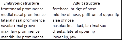
Below is a summary of the contributions of the prominences to the adult face:

table5

  • median cleft lip (incomplete fusion of medial nasal prominences)

  • frontonasal dysplasia (hyperplasia of inferior frontonasal prominence, thus preventing fusion of the medial nasal prominences)

  • forms via the fusion of the two medial nasal prominences in the midline (of course, this midline fusion is driven via growth of the maxillary prominences which pushes the nasal prominences toward to the middle)

  • consists of the premaxillary segment of the maxilla, which contains the four incisors and the incisive canal


Complete fusion of the primary and secondary palate is a complex process involving growth of the component tissues, epithelial to mesenchymal transformation, cell migration, and programmed cell death at fusion sites –disruption of any part of this process can result in cleft palate.  Given the involvement of the maxillary and nasal prominences, cleft palate is often (but NOT always) accompanied by cleft lip.

ANSWER


  1. overgrowth of the frontonasal process

  2. incomplete fusion of the frontonasal and maxillary processes

3.  The condition shown in the figure below was most likely caused by:

A.  1st pharyngeal pouch.
B.  2nd pharngeal pouch.
C.  ventral portion of the 3rd pharyngeal pouch.
D.  dorsal portion of the 4th pharyngeal pouch.
E.  foramen cecum at the base of the tongue.

ANSWER

ANSWER


Questions 7-10 refer to the list below.  Select the most appropriate structure in the list for each of the following statements or descriptions (each labeled structure may be used once, more than once, in combination with other structures, or not at all).  If a statement or description refers to a structure NOT in the list then the correct answer would be "NONE of the above."

8.  gives rise to the mandible

ANSWER

10.  its derivatives are innervated by the hypoglossal nerve

ANSWER


11.  innervated by the facial nerve  


13.  innervated by cranial nerve VIII 


ANSWER


17. Which of the following could you expect to find upon further examination?


You are viewing 1/3rd of the document.Purchase the document to get full access instantly

Immediately available after payment
Both online and downloadable
No strings attached
How It Works
Login account
Login Your Account
Place in cart
Add to Cart
send in the money
Make payment
Document download
Download File
img

Uploaded by : Breno Novaes

PageId: DOC0EDCE37