
Form-ing the muscular interventricular septum fig


180Part IISystems-Based Embryology
| Aorta | Pulmonary valves |
|---|
Tricuspid orifice
Moderator band
|
|---|
Outflow channel
of right ventricle
Neural tube
Dorsal
aorta
Septum Formation in the Ventricles
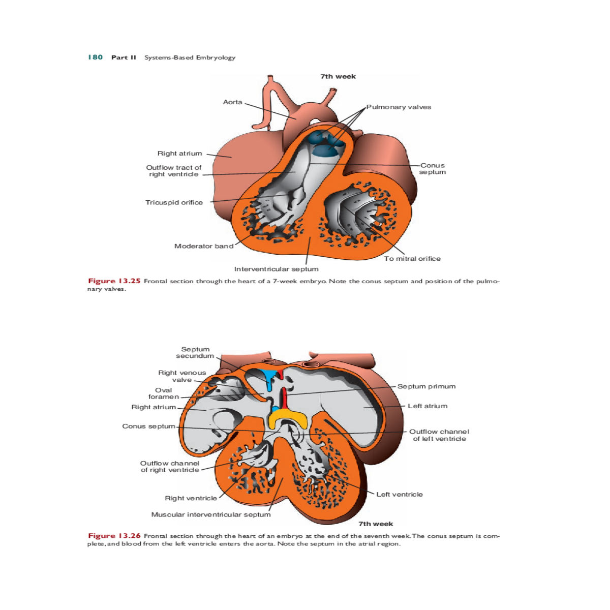
By the end of the fourth week, the two primitive ventricles begin to expand. This is accomplished by continuous growth of the myocardium on the outside and continuous diverticulation and trabecula formation on the inside (Figs. 13.19 and 13.26).
| Minor truncus swelling | Aorta | |
|---|---|---|
|
Right
truncus
swelling


