

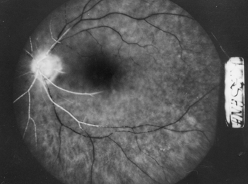
Central retinal artery occlusion crao fig initial stages
602
 |
 |
|---|
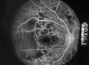
| Fig.1: Severe NPDR- with ischemic | |||
|---|---|---|---|
| maculopathy. Extensive CNP areas are seen. | |||
| Hypofluorescence of non-perfusion areas is outlined by capillaries unlike hypofluorescence | |||
|
|||
|
|||
| v) | Dye usually stains and leaks from the venous wall in | i) |
|
|
|||
| ii) | |||
9. Non-ischemic Central retinal venous occlusion (CRVO) - FFA advisable only after hemorrhages resolve significantly i.e. after 3-4 months.
i) Delayed central retinal venous filling (normally in 20- 25 seconds) and also delayed emptying.
ii) Engorged and tortuous retinal veins FFA picture depends on whether retina, choroid or
iii) Retinal hges (superficial) block both retinal & choroidal optic nerve is involved.
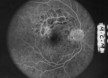
vii) Macula may show diffuse leaking from perifoveal
capillaries (may by cystoid with typically petalloid
ii) Extensive CNP areas all over the fundus.
ii) Cotton wool spots are seen as CNP areas (central hypofluorescence bordered by dilated capillaries).
| v) | ||
|---|---|---|
|
|
|
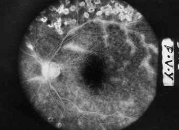
13. Central Retinal Artery Occlusion (CRAO) (Fig.4)
| characteristics. Retinal hemorrhages tend to be in mid | i) |
|
|
|---|---|---|---|
| 561 | February, 2006 | ||
ii) Markedly delayed arm retinal may even mask the macroaneurysm. vi) After about 2 weeks although dye is broken (Box-carring). involved segments.
iii) Hypofluorescent areas also occur due to blocked vii) In combined CRVO & CRAO, filling of arteries by dye |
|---|
 17. Purtscher’s
retinopathy-
17. Purtscher’s
retinopathy-14. Branch retinal arterial occlusion (BRAO)-
Picture on FFA is similar to CRAO except that it is confined to the affected segment.
i) Hard drusen show as early hyperfluorescent dots increasing in AV phase but fading in later phases.
|
ii) | |
|---|---|---|
iii) Focal hyperpigmentation and RPE atrophy- Mottled hyperfluorescence i.e. blocked background choroidal fluorescence interspersed with window defects.
|
Telangiectatic and aneurysmal vessels are well made |
|---|
iv) Vascular abnormalities as seen on FFA are much more than that seen on fundus examination.
16. Retinal arterial macroaneurysm-
� Early window defects
� Hypofluorescent areas due to choriocapillaris dropouts.
i) Classical CNVM- is well outlined even in early phases as hyperfluorescent patch of lacy pattern with increase in intensity and size in mid and later phases.
ii) Occult CNVM shows 2 patterns on FFA -
| 562 | DOS Times - Vol. 11, No. 8 |
|---|


