The Lower Limb Assignment Help
The bones of appendicular skeleton that includes leg and foot forms the lower limb. Also the bones of pelvic girdle makes the part of lower limb.
Looking for lower limb Assignment Help, call our online tutors and get the further details.
Pelvic girdle
Pelvic girdle is ring like structure that connects axial skeleton to lower limb. It consists of two hip bones named as sacrum and coccyx. The superior part of the pelvic is termed as greater pelvic and the inferior part is called lesser pelvic. The connections between them is called pelvic inlet. Pelvic girdle has four attachment, namely Sacroiliac joint (between ileum and sacrum), Sacrococcygeal symphysis (between sacrum and coccyx), Pubic symphysis (between pubic body of the two hip bones). Further two hipbones are major part of pelvic girdle that in turn introduces three connections, sacroiliac joint (with sacrum), pubis symphysis (with equivalent hipbone) and hip joint (with head of femur).
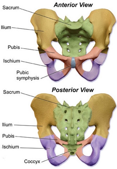
The hip bone
The three important parts of hip bone are: ilium, pubis and ischium. The fusion of these parts forms a cup-shaped socket called acetabulum.
Illeum
- It is broadest of the all three parts.
- Just above the acetabulum, ileum expands to form wing like structure that give this bone broader shape.
- The inner portion of this wing is concave in shape and called iliac fossa because it provides attachment for iliacus muscle and the external portion is convex and named as gluteal surface which serves as attachment for gluteal muscle.
- Iliac crest is formed at the thickened portion of superior margin.
Pubis
- It is located at the anterior portion of the hip bone.
- It is divided into body and two branches namely superior rami and inferior rami.
- The body is medially located.
- The superior rami forms part of acetabulum by extending laterally from the body.
- The inferior rami joins acetabulum.
- Obturator foramen is enclosed in between superior and inferior rami.
The ischium
- Ischium makes up the posterioinferior part of hip bone.
- Greater sciatic notch is present at the posterior side of the ischium.
- The combination of inferior ischial ramus and inferior pubic ramus forms ischiopubic ramus.
- Sacrospinous ligament and sacrotuberous ligament are attach to ischium.
Sacrum:
- It forms posterior part of the pelvis.
- It is thick bone formed by the fusion of 5 sacral vertebrae.
- The bones of sacrum includes base, apex and four surfaces.
- Base connects with fifth lumbar vertebrae superiorly.
- Apex touches coccyx at the inferior side.
- Auricular surfaces are located laterally on the sacrum and each one of them articulate with auricular surface of ileum.
- Coarse dorsal surface and smooth pelvic surface are two surfaces of sacrum.
Coccyx
- It is the terminal part of vertebral column.
- It consists of apex, base, anterior and posterior surface and two lateral surfaces.
- Base is superiorly located and has surface for articulation with the sacrum.
- Apex is inferiorly located.
- Sacrococcygeal joint is formed by the articulation coccyx with symphysis.
- 5 ligaments namely anterior Sacrococcygeal ligament, deep posterior Sacrococcygeal ligament, superficial posterior Sacrococcygeal ligament, lateral Sacrococcygeal ligaments and interarticular ligaments supports sacrococcygeal.
- Gluteus Maximus helps in attachment of coccyx.
Want to get your anatomy assignment rechecked? Email us at support@assignmenthelp.net to opt our service of lower limb Assignment Help.
Femur (Bone of leg)
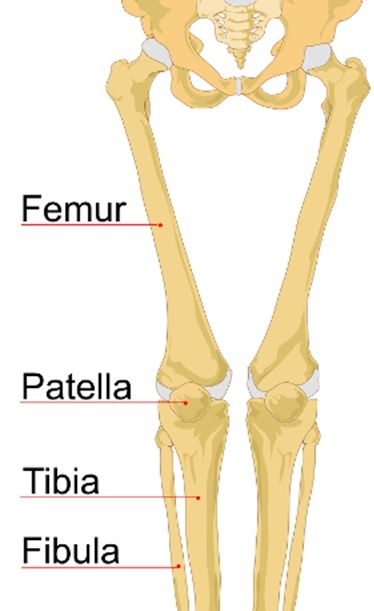
- It is a long bone of leg.
- Divided into three areas, proximal, shaft and distal.
- Proximal area forms the hip joint in combination with pelvis.
- Proximal area consist of head, neck, trochanter and bony ridges.
- Head is a smooth surface that articulates with acetabulum of pelvis at the hip joint.
- Neck connects head with the shaft and projects superiorly and medially.
- Trochanter is divided into greater trochanter (originates from anterior shaft) and lesser trochanter (originates from posteriomedial side).
- Two bony ridges of proximal part are intertrochanteric line (runs on the anterior surface of femur) and intertrochanteric crest (runs on the posterior surface of femur).
- Shaft of femur descends slightly in medial direction.
- The distal area has condyles at the medial end and lateral end.
- These condyles connect with tibia and patella to form knee joint.
Patella (Bones of knee)
- Present at the front of knee joint.
- Superiorly it attaches to quadriceps and inferiorly it attaches to patellar ligament.
- It is triangular shaped with anterior and posterior regions.
- Patella is divided into apex and base.
- Apex is inferiorly situated and connected to tibial tuberosity.
- Base forms the superior part of the bone and allows attachment for quadriceps tendon.
Tibia
- It is another main bone of the leg and second largest bone in the body.
- The proximal part of the tibia articulate with knee and the distal part of tibia articulate with ankle.
- The proximal part of tibia is widened due to two condyles, medial and lateral condyles.
- Intercondylar eminence is present in between these condyles.
- Tibial tuberosity is present at the anterior part of proximal tibia.
- The shaft of tibia consist of three regions; anterior border (marked by tibial tuberosity), posterior border (marked by soleal line) and lateral border.
- The distal end is widened. Lateral side of distal end has notch known as fibular notch that binds tibia with fibula.
Looking for exact and accurate information on skeletal system? Check our online lower limb Assignment Help service and get the details.
Fibula
- In combination with tibia, it makes the bone of leg.
- It is thinner and present laterally to tibia.
- The proximal end of fibula provide attachment for lateral condyle of tibia.
- Shaft is divided into anterior, lateral and posterior surface.
- The lateral surface of distal area in fibula continues inferiorly and is called lateral malleolus.
Tarsals (Bones of foot)
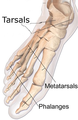
- These are seven unequal shaped bones present in the proximal area of the foot.
- Tarsals are divided into proximal, intermediate and distal portion.
- The proximal part consist of talus and calcaneus bones, talus being the superior and calcaneus present at the underneath of talus.
- Talus articulate superiorly with tibia and fibula to form ankle joint, inferiorly it forms subtalar joint and anteriorly it forms talonavicular joint.
- Calcaneus forms subtalar joint at the superior portion and calcaneocuboid joint at the anterior portion.
- At the intermediate position, tarsal contain one bone named navicular that articulate with talus at the posterior side and cuneiform at the anterior side and cuboid bone at the lateral side.
- At the distal portion, a cuboid and three cuneiforms are present that connect with metatarsals of foot.
Metatarsals (Bones of foot)
- Located between tarsals and phalanges.
- Each metatarsals has head distally situated, base proximally situated and shaft bone that join proximal and distal portion.
- At the proximal portion, tarsometatarsal joint is formed by articulation of metatarsal bases and cuneiforms.
- Intermetatarsals is formed laterally by the articulation of adjacent metatarsals.
- Metatarsophalangeal joint is formed distally due to articulation of metatarsal head with proximal phalanx.
Phalanges (Bones of foot)
- These are the bones of toes.
- There are three portions, proximal, distal and intermediate.
- Only the main toe has proximal and distal parts.
Want the help of experts to help you write your lower limb assignment? Get in live chat with our biology team for anatomy Assignment Help and get field specific biology tutor to guide you in writing your skeletal system assignment.
Image Reference: https://commons.wikimedia.org/