Muscles Of Anterior Compartment Of The Thigh Assignment Help
In the anterior compartment of the thigh, there are four major muscles; the pectineus, lliopsoas, Sartorius, quadriceps femoris.
Pectineous
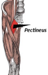
It is flat in shape forming the base of the femoral triangle. The dual innervation in this muscle makes it a transitional muscle between the compartment of anterior thigh and medial thigh. Originating from pectin pubis, this muscle gets attach to the pectineal line on the posterior part of the femur. It has a nerve supply of femoral nerve. The main function of pectineous muscle is flexion and adduction at the hip joint.
Facing difficulty in writing lengthy assignment on muscles of anterior compartment of thigh? Seek the help from our tutors.
Illiopsoas:
Due to the close relationship between iliacus and psoas major muscles of hip, they are jointly called Illiopsoas. Though the origin of both muscles are different, iliacus arises from ilium and psoas major arises from the lumbar vertebrae, but a common insertion at lesser trochanter of femur gives a common name for both these muscles. Therefore, illiopsoas help in the flexon of the hip that allows to perform number of activities.
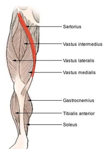
Sartorius:
It is elongated, thin and band like muscle in the thigh. It runs across thigh in inferomedial direction. It originates from anterior superior iliac spine descending obliquely across the hip joint. It attaches to the superior medial surface of the tibia. It has a nerve supply of femoral nerve.
Muscles Of Anterior Compartment Of The Thigh Assignment Help By Online Tutoring and Guided Sessions at AssignmentHelp.Net
Wanting anatomy Assignment Help from professional tutors? Call on the given number or email us for instant help.
Quadriceps femoris:
Four individual muscles namely three vastus muscles and the rectus femoris makes up quadriceps femoris.
Vastus muscles:
This is of three types: vastus medialis, vastus intermedius and vastus laterlis. Vastus medialis muscle is present at the sides and front of the thigh and is a primary extensor of knee. Likewise, vastus laterlis is present on the lateral side of the thigh and acts as an extensor of leg at the knee. It arises from common origin on the trochanter of femur. Also the muscle fibers of this muscle extend anteriorly and distally to merge with rest three muscles. Vastus intermedius lies deep to other muscles.
Rectus femoris
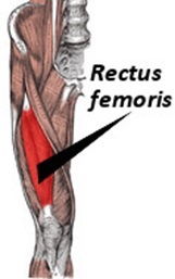
It originates from ilium and runs straight down the leg therefore attaching to the patella by quadriceps femoris tendon. It is supplied by femoral nerve. This is the only muscle that flexes at hip joint and extends at the knee joint.
| Muscle | Origin | Insertion | Nerve Supply | Nerve Roots | Action |
|---|---|---|---|---|---|
| Sartorius | Anterior superior iliac spine | Upper medial surface of shaft of tibia | Femoral nerve | L2,3 | Flexes, abducts, laterally rotates thigh at hip joint;flex & medially rotates leg at knee joints |
| Iliacus | Iliac fossa of hip bone | With psoas into lesser trochanter of femur | Femoral nerve | L2,3 | Flexes thigh on trunk |
| Psoas | Transverse processes, bodies, & intervertibral disc of the 12th thoracic & five lumber vertebrae | With iliacus into lesser thochanter of femur | Lumber plexus | L1,2,3 | Flexes thigh on trunk |
| Rectus demoris | Anterior inferior iliac spine & ilium above acetabulum | Quadriceps tendon into patella, then via ligamentum patellae into tubercle of tibia | Femoral nerve | L2,3,4 | Extensor of leg at knee joint; flexes thigh at hip joint |
| Vastus medialis | Upper end and shaft of femur | Quadriceps tendon into patella, then via ligamentum patellae into tubercle of tibia | Femoral nerve | L2,3,4 | Extension of leg at knee joints |
| Vastus laterali8s | Upper end and shaft of femur | Quadriceps tendon into patella, then via ligamentum patellae into tubercle of tibia | Femoral nerve | L2,3,4 | Extension of leg at knee joints |
| Vastus intermedius | Anterior & lateral surface of femur | Quadriceps tendon into patella, then via ligamentum patellae into tubercle of tibia | Femoral nerve | L2,3,4 | Extension of leg at knee joint. |
If you require anatomy Assignment Help in muscular system, contact assignmenthelp.net and receive help from the professionals.
Image Reference: commons.wikimedia.org