Muscles connecting The Upper Limb To The Thoracic Wall Assignment Help
There are four important muscles that connects upper limb with the thoracic wall, namely Pectoralis major, Pectoralis minor, Subclavius and Serratus anterior.
Pectoralis major:
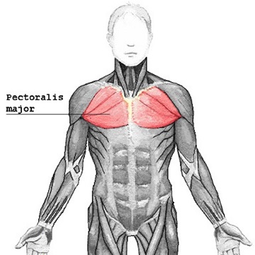
It is a large muscle present in the upper limb of human skeletal system. This muscle is fan shaped and makes most portion of the anterior wall of axilla. It originates from clavicle, sternum and upper six coastal cartilage. The two head of this muscle are named as clavicular head and sternocostal head that are attached to clavicle and sternum respectively. Both these heads can either work in combination or independently to allow the rotation of humerus. The pectoralis major is supplied with two nerves derived from brachial plexus called medial pectoralis nerve and lateral pectoralis nerve.
Looking for anatomy Assignment Help? Call or email us to receive desired assistance.
Pectoralis minor:
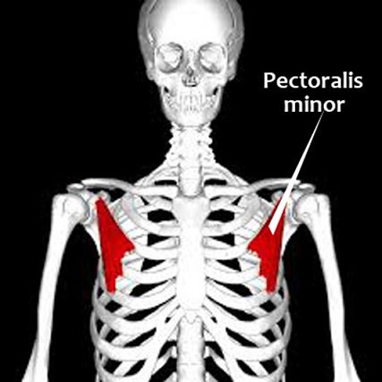
The size of pectoralis minor is smaller in comparison to pectoralis major. It is triangular shape that patents from anterior part of third, fourth and fifth ribs. It then insert to corocoid process of scapula. Pectoralis minor lies underneath to pectoralis minor and together they makes the anterior wall of axilla. The contraction of this muscle generates inferior motion. It is supplied by medial pectoral nerve that comes from brachial plexus.
Serratus anterior:
Muscles connecting The Upper Limb To The Thoracic Wall Assignment Help By Online Tutoring and Guided Sessions at AssignmentHelp.Net
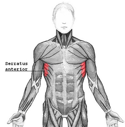
It originates from upper eight ribs and inserts into medial border and interior angle of scapula. It is displaced more laterally in the chest forming a medial wall of axilla. The main function of serratus anterior is to allow rotation of scapula. Also serratus anterior holds scapula against the ribcage. It is supplied by long thoracic nerve.
Are you looking for Muscular system Assignment Help? If so contact our online Course Helper to guide you in the provided task.
Subclavicus:
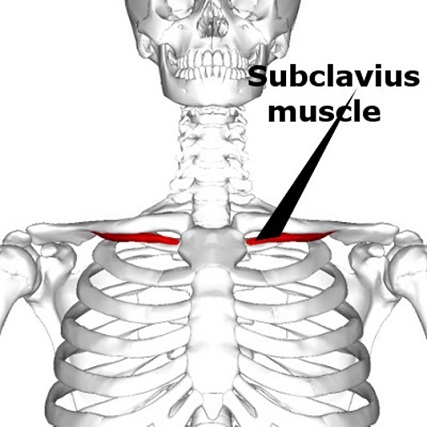
It is a minor muscle that is located deep inside the chest. These muscle is two in number that inserts into inferior side of clavicle bone, specifically called collar bone. It originates from first costal cartilage and inserts into clavicle. It is supplied by the nerve from upper trunk of the brachial plexus. Therefore, during the movement of shoulder girdle it depresses the clavicle.
| MUSCLES | ORIGIN | INSERTION | NERVE SUPPLY | NERVE ROOTS | ACTION |
|---|---|---|---|---|---|
| Pectoralis major | Clavicle,sternum, and upper six costal cartilage | Lateral lip of bicipital groove of humerus | Medial and lateral pectoral nerve from brachial plexus | C5,6,7,8;T1 | Adducts arm and rotates it medially;clavicular fibres also flex arm |
| Pectoralis minor | Third, fourth, and fifth ribs | Coracoid process of scapula | Medial pectoral nerve | C6,7,8 | Depresse point of shoulder, and elevates the ribs |
| Subclavius | First costal cartilage | Clavicle | Nerve to sub clavius | C5,6 | Depresses clavicle |
| Serratus anterior | Upper eight ribs | Medial border and inferior angle of scapula | Long thoracic nerve | C5,6,7 | Draws the scapulla forward and rotates scapula. |
Hence these are four important muscles that connects upper limb to the thoracic wall. If you want to obtain detailed information about the muscles connecting upper limb to the thoracic wall, seek the assistance of our online tutors.
At assignmenthelp.net, you can obtain help to receive information on human system, all its parts, joints, nerves and the functions. If you want anatomy Assignment Help or anatomy Assignment Help, even then you can contact our tutors for required help.
Image reference: en.wikipedia.org, commons.wikimedia.org