Muscle of Medial Fascial Compartment Of The Thigh Assignment Help
The area between pelvis and the knee is termed as thigh. Thigh has a single bone called femur and is mainly covered by five important muscles, namely: Sarsotius muscle, Quadriceps femoris muscle, Adductor longus muscle, Hamstring muscle and femoral triange. Thigh is grouped into three major compartments, called anterior, posterior and medial compartment and all these three compartments are separated by fascia.
Focusing on the main topic of this article, there are five important muscles in the medial compartment of the thigh, namely; gracilis, Pectineus, adductor brevis, adductor longus and adductor magus. These muscles of medial compartment are together called hip adductors that adduct the thigh. Medial compartment of thigh is supplied by obturator nerve L2, 3, 4 and the arterial supply is through obturator artery.

Looking for Assignment Help on the topic of muscle of medial compartment of the thigh? Get assistance from online tutors at assignmenthelp.net.
Abductor longus
- It is big and flat in shape.
- Originates from medial portion of superior pubis ramus.
- It forms the medial border of femoral triangle and expands into a fan shape therefore inserting to linea aspera in femur.
- Posteriorly it is connected by adductor brevis and adductor magnus and anteriorly with the fascia lata.
- It is innervated by obturator nerve.
- Helps in adduction and medial turning of thigh.
Muscle of Medial Fascial Compartment Of The Thigh Assignment Help By Online Tutoring and Guided Sessions at AssignmentHelp.Net
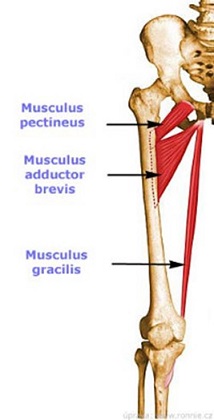
Adductor brevis
- This muscle is short and lies beneath the adductor lungus.
- It originates from inferior pubis ramus.
- This muscle inserts into aspera of femur.
- Anteriorly it is connected with pectineus and adductor longus and posteriorly with adductor magnus and at the outer border with the obturator externus.
- It is innervated by obturator nerve.
- Helps in Adduction of the thigh.
To get anatomy Assignment Help on topic of muscular system, get in touch with the team of learned and educated tutors, email at support@assignmenthelp.net.
Gracilis
- It is the most superficial muscle of the medial compartment.
- It originates from pubis symphysis and inferior pubis ramus.
- This muscle inserts into medial tibia.
- Internally it is in relation with the fascia lata and exteriorly it is connected to adductor longus, brevis and magnus.
- It is innervated by obturator nerve.
- Helps in adduction of thigh and flexon of leg.
Pectineus
- It is flat muscle, quadrangular in shape.
- It originates from pubis.
- This muscle inserts into lesser trochanter.
- It is connected to fascia lata at the anterior surface, posteriorly with capsule of hip joint, obturator externus and adductor brevis, externally with psoas major and internally with outer edge of adductor longus.
- It is supplied by femoral nerve.
- Helps in adduction, hip flexon and medial rotation.
Adductor magnus
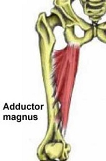
- It is largest muscle of medial compartment.
- Adductor magnus is further classified into two parts namely: adductor part and hamstring part.
- Adductor part patents from inferior rami of pubis and the rami of ischium which then attaches to aspera of the femur.
- The hamstring originates from ischial tuberosity and then attaches to adductor tubercle on distal and medial side of femur.
- Anteriorly it is connected pectineus, adductor brevis and adductor longus, posteriorly with semitendinosus, semimembranosus, biceps and gluteus maximum muscle. Internally it is linked with gracilis and Sartorius and by its upper border with obturator externus and quadratus femoris.
- Adductor part is supplied by obturator nerve and hamstring part is supplied by tibial nerve.
- Both these parts help in flexing and extending the thigh.
If you are not able to get detailed and organized information for your topic of anatomy assignment, get in touch with the tutors of assignmenthelp.net. They will help you in completing your assignment before the provided deadline, thus including relevant pictures in your work.
Image references:
http://memorize.com/adductor-longus/kms013
http://medicina.ronnie.cz/img/data/clanky/normal/1862_1.jpg
http://www.sportsinjuryclinic.net/media/Hip_and_groin/adductor-magnus185.jpg
| MUSCLE | ORIGIN | INSERTION | NERVE SUPPLY | NERVE ROOTS | ACTION |
| GRACILIS | Inferior ramus of pubis, ramus of ischium | Upper part of shaft of tibia on medial surface | Obturator nerve | L2,3 | Adducts thigh at hip joints ;flex leg at knee joint |
| Adductor longus | Body of pubis, medial to pubic tubercle | Posterior surface of saft of femur | Obturator nerve | L2,3,4 | Adducts thigh at hip joint & assist in lateral rotation |
| Adductor bravis | Inferior ramus of pubis | Posterior surface of saft of femur | Obturator nerve | L2,3,4 | Adducts thigh at hip joint & assist in lateral rotation |
| Adductor magnus | Inferior ramus of pubis,ramus of ischium, ischial tuberosity | Posterior surface of saft of femur | Obturator nerve & sciatic nerve | L2,3,4 | Adducts thigh at hip joint & assist in lateral rotation |
| Pectineus | Superior ramus of pubis | Upper end of linea aspra of shaft of femur | Femoral nerve | L2,3 | Flexes and adducts thigh at hip joint |
Email Based homework Help in Muscle of Medial Fascial Compartment Of The Thigh
To submit Muscle of Medial Fascial Compartment Of The Thigh assignment Upload Assignment.