Bones of Shoulder Girdle and Arm Assignment Help
Shoulder girdle gives strength to the arm and helps in diverse range of gesture. This is the reason it is made of complex arrangement of bones, muscles and ligaments. The three important bones that make up shoulder girdle are clavicle, scapula and humerus. Know more about clavicle, scapula and humerus here.
Therefore, anatomically the movement of shoulder girdle is possible due to three important joints named as Glenohumeral joint, Acromioclavicular joint and Sternoclavicular joint. The point where two or more bones meet is termed as joint. Beside these anatomical joints, two physiological joints also take part in movement of shoulder girdle named as suprahumeral joint and scapulocostal joint. In this article, you will be presented only an overview of three anatomically important joints of skeletal system.
In need of anatomy Assignment Help on skeletal system? Contact assignmenthelp.net to get help with the desired section.
Joints of shoulder girdle
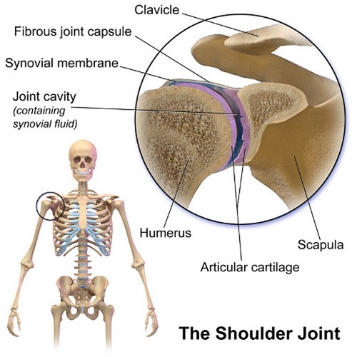
Glenohumeral joint:
It is an important joint that links humerus with the thorax. GH joint is ball and socket like joint that is formed by the articulation between the head part of humerus and cup like depression of scapula called as glenoid fossa. This articulated surface is covered by hyaline cartilage. The larger head of humerus than the glenoid fossa can possibly result in inherited stability of this joint, so this is the reason fibrocartilage rim called glenoid labrum is present to reduce this disproportionate in the surface. Extending from anatomical neck of the humerus to the border of glenoid fossa, a joint capsule is present. It is a fibrous sheath that encloses the structures of the joint, hence allowing greater mobility. Also the friction between articular surfaces is reduced by the presence of synovial fluid produced by the inner lining of synovial membrane.
The friction that is present in the shoulder joint is reduced by synovial bursae that acts as cushion between tendons and other joint structures. Moreover together with ligaments namely Glenohumeral ligament, Coroacohumeral ligament and Transverse humeral ligament, shoulder girdle permits different types of movement including extension, flexion, abduction, adduction, lateral rotation and medial rotation. Therefore, maximum motion in the shoulder girdle is provided by GH joint. The movement of arm in desired direction is also possible due to GH joint present in the shoulder girdle.
Sternoclavicular joint
It is the joint between clavicle and manubrium of the sternum. Therefore the sternal end of clavicle, the manubrium of sternum and part of first costal cartilage makes of Sternoclavicular joint. All these articulated surfaces are covered by fibrocartilage and separated into two compartments by fibrocartilaginous articular disc. Also the joint shell present in sternoclavicular joint consists of outer layer that is fibrous and inner synovial membrane. Extending from the epiphysis of the sternal end of clavicle to the borders of articular surface and the articular disc is fibrous layer whereas present in the inner surface is synovial membrane that produces synovial fluid. Synovial fluid reduces friction between articular surfaces. Therefore, the major ligaments namely Sternoclavicular ligaments, Interclavicular ligament and Costoclavicular ligament promotes stability of this joint. Therefore, sternoclavicular joint allows elevation, depression, protraction, retraction and rotation movements of the shoulder girdle.
Acromioclavicular joint
The point of articulation between lateral ends of clavicle with acromion of scapula is called acromioclavicular joint. The main identification of acromioclavicular joint is that the articulated surface are lined with fibrocartilage and the joint cavity is divided by an articular disc.
The two articular surface of joint shell of acromioclavicular joint consist of loose fibrous layer that gives rise to articular disc. Also the fibers from a muscle namely trapezius muscle covers the posterior part of joint shell. Moreover, the synovial membrane present in the inner side release synovial fluid that lines the joint. Along with acromioclavicular joint, conoid and trapezoid ligaments promotes axial and anteroposterior movement.
Bones of Shoulder Girdle and Arm Assignment Help By Online Tutoring and Guided Sessions from AssignmentHelp.Net
Looking for shoulder girdle Assignment Help, take the assistance from our Biology experts.
Joints of arm
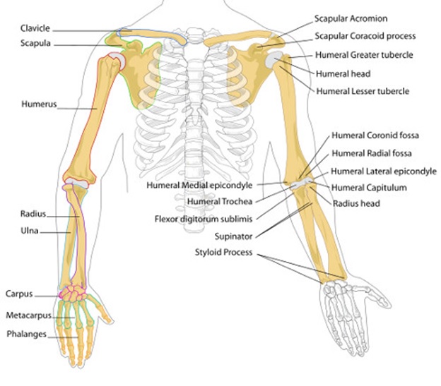
To read about bones of arm, click here. The main arm is connected to the forearm at the point known as elbow, which is a part of upper limb. The bones at the upper limb of arm are epicondyles along with olecranon process. Therefore the joint here is called synovial joint or hinge joint.
Trochlear notch in the ulna articulate with trochlear notch in humerus and head of humerus articulate with capitulum of humerus thus making elbow joint also called pivot joint. This joint is enclosed inside a capsule hence allowing extension and flexion of arm. Radial collateral ligament and ulnar collateral ligament helps in stability of this joint.
Radioulnar joint
It is formed by articulation of radius and ulna at two points. When the head of radius articulate with radial notch of ulna, proximal radioulnar joint is formed. Similarly, when the ulnar notch of radius articulate with ulnar head, distal radioulnar joint is formed. Therefore, proximal radioulnar joint and distal radioulnar joint together makes up radioulnar joint. Interosseous membrane is present in between radius and ulna that holds them together and transfer force from radius to ulna.
Wrist joint
It is the joint between forearm and the hand. It is distally formed by proximal rows of carpal bones (except pisiform) and proximally formed by distal end of the radius with the articular disk. The joint shell of wrist joint is two layered. Outer layer attaches to ulna and carpal bones (proximal row) whereas the internal layer is made of synovial membrane that secretes synovial fluid. Palmar radio carpal, dorsal radio carpal, ulnar collateral and radial collateral are major ligaments in the wrist joint that helps in its stability. The movement of wrist joint include flexion, extension, adduction and abduction.